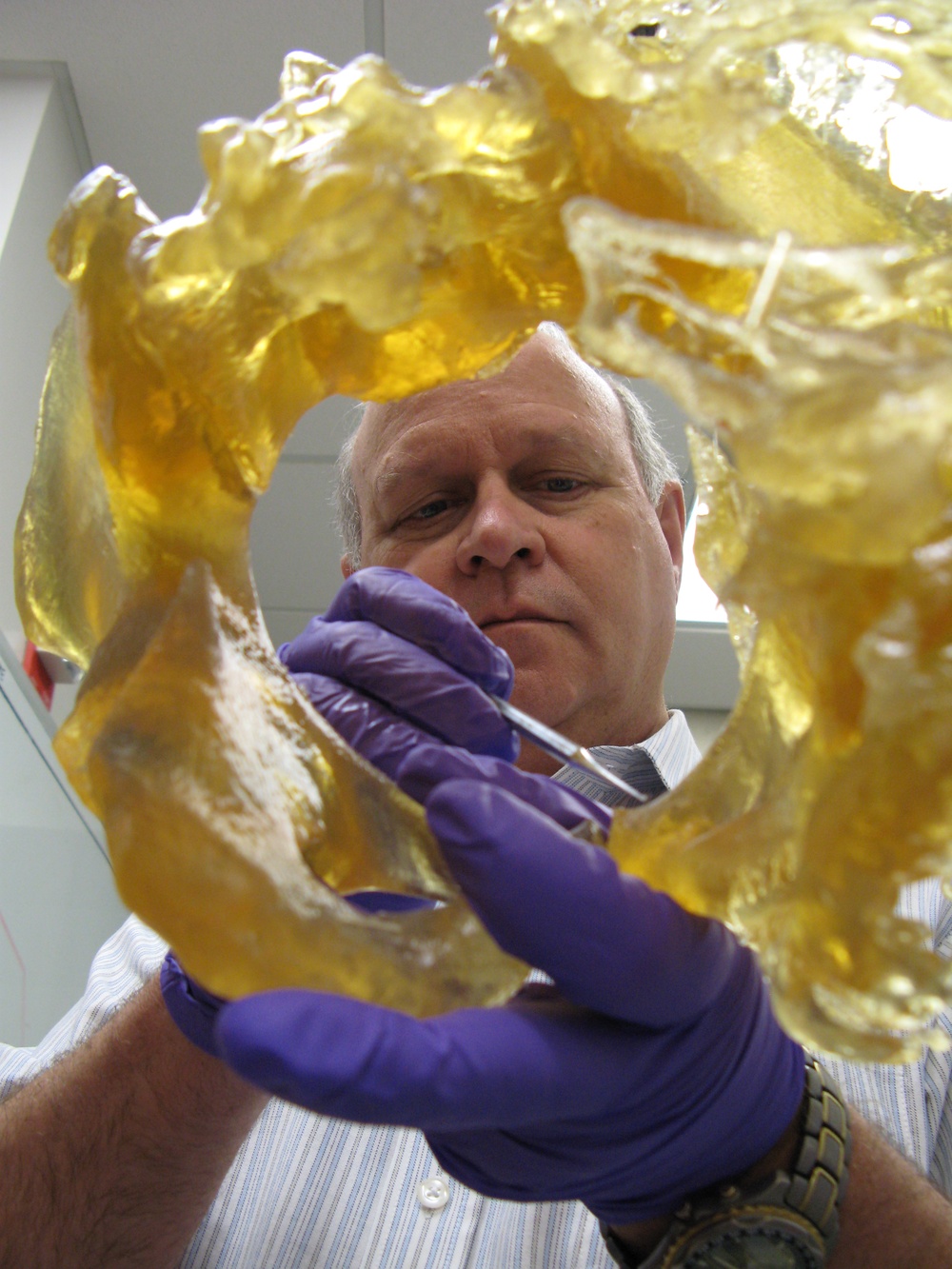
Dr. Stephen Rouse, a retired military dentist who runs Walter Reed Army Medical Center’s 3-D Medical Applications Center, cleans a medical model of a pelvis with heterotopic ossification, or HO. The growth is random and can form a lacy network of new bone under the skin, similar in appearance to coral formations, causing problems for amputees, especially when fitting and wearing prosthetics. The bony growth sometimes encapsulates major arteries or nerves, making surgery risky. A 3-D model gives surgeons and patients a clearer look at problems before operating. Photo by Fred W. Baker III
| Date Taken: | 04.23.2007 |
| Date Posted: | 07.03.2025 22:30 |
| Photo ID: | 9157981 |
| VIRIN: | 070423-D-D0439-8598 |
| Resolution: | 2736x3648 |
| Size: | 3.85 MB |
| Location: | (UNDISCLOSED LOCATION) |
| Web Views: | 1 |
| Downloads: | 0 |

This work, 3-D Imaging Helps Surgeons Tackle Challenging Bone Growth in Amputees [Image 4 of 4], must comply with the restrictions shown on https://www.dvidshub.net/about/copyright.